선천성 귀 기형, Congenital malformations of ears
| 선천성 귀 기형의 종류 |
1. 소이증 smaller ear
귓바퀴가 비정상적으로 작은 선천성 귀 기형
2. 대이증 Macrotia
귓바퀴가 비정상적으로 큰 선천성 귀 기형
3. 무이증 No auricle
귓바퀴가 없는 선천성 귀 기형.
4. 대이 Auriclar skin tag
외이도 입구 바로 앞 부위, 이주 바로 앞 부위에 쌀알 크기만 한 피부 주머니, 또는 콩알 크기만 한 피부 주머니. (사진2 참조)
5. 이루공 Cogenital auricular fistula
외이도 입구의 바로 앞 부위(이주 바로 앞 부위)에 있는 안면 피부에 바늘구멍 크기만 한 구멍이 선천적으로 뚫려 있는 귀 기형(사진 28, 29 참조).
6. 부이 Accessory ear
외이도 입구의 바로 앞 부위(이주 바로 앞 부위)에 있는 안면 피부에 두드러진 피부돌기가 선천적으로 나 있는 귀 기형(사진 22~27 참조).
부이는 선천성 귀 기형 중 가장 흔한 귀 기형이다.
7. 선천성 무 귀 Cogenital anotia
외이도와 중이 등이 선천적으로 없는 귀 기형.
8. 돌출 귓바퀴 Protrude auricle
사진 18 참조.
9. 불완전하게 생긴 이륜 Incomplete auricle
사진 19 참조.
10. 늘어진 귓바퀴 Floppy auricle
사진 20 참조.
11. 이륜(귓바퀴)에 있는 달윈 돌기 Darwin ear point.
사진 21 참조.
- Microtia: Underdeveloped outer ear
- Anotia: Missing one or both ears
- Protruding ears: More than 2 cm from the head
- Constricted ears (cup ears): Flattened or rolled outer ears
- Cryptotia: Upper ear underneath the scalp skin
- Stahl’s ear: Pointed outer ear
- Ear tag (accessory tragus): Cartilage bump in front of the ear
- Congenital earlobe deformities: Duplicate earlobes or earlobes with clefts or skin tags
- Ear hemangiomas: Small to large benign tumors
| 선천성 귀 기형의 치료 및 합병증 |
- 대이가 박테리아에 감염되면 대이가 곪을 수 있다.
- 적절한 항생제로 치료해야 한다. 대이가 자주 곪으면 대이를 수술로 제거 치료해 주기도 한다.
- 대이가 곪지도 않고 미관상 큰 문제가 되지 않으면 그대로 두고 관찰 치료를 한다.
- 이루공도 특별히 치료해 줄 것 없이 그대로 두고 보는 것이 보통이다. 때로는 감염될 수 있다.
- 부이가 가늘고 길쭉하게 생겼을 때는 부이 맨 밑 부분을 실로 꼭 매어 부이를 떼 낼 수 있다(사진 23 참조).
- 사진 23에서 볼 수 있는 것과 같이 피부에 가늘게 붙어 있는 부이 맨 밑 부분을 실로 꼭 잡아맨 후 1~2주 되면 부이가 괴사되고 그 부이 뿌리가 피부에서 자연적으로 떨어지는 것이 보통이다. 그렇지 않으면 수술로 절제하여 떼 낼 수 있다.
- 외이나 외이도, 또는 중이가 태어날 때부터 없으면 외이나 외이도를 성형수술로 새로 만들어 줄 수 있다.
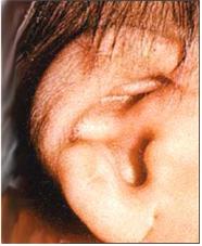
사진18. 돌출 귓바퀴
출처; Used with permission from Mead Johnson Nutrituional Division과 소아가정간호백과
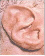
사진 19. 불완전하게 생긴 이개
출처; Used with permission from Mead Johnson Nutrituional Division과 소아가정간호백과
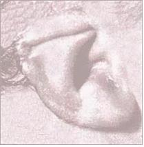
사진 20. 늘어진 귓바퀴
출처; Used with permission from Mead Johnson Nutrituional Division과 소아가정간호백과
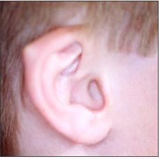
사진 21. 이륜(귓바퀴)에 있는 달윈 돌기
Copyright ⓒ 2012 John Sangwon Lee, MD., FAAP
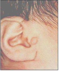
사진 22. 외이도 앞 부위에 난 부이
Copyright ⓒ 2012 John Sangwon Lee, MD., FAAP
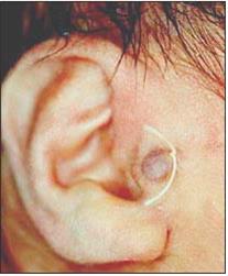
사진 23. 부이의 맨 밑 부분을 실로 꼭 매어 2주 지나면 부이가 괴사된 후 떨어질 수 있다.
Copyright ⓒ 2012 John Sangwon Lee, MD., FAAP
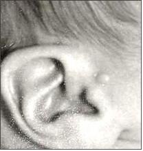
사진 24. 외이도 앞 부위에 난 부이
Copyright ⓒ 2012 John Sangwon Lee, MD., FAAP
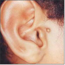
사진 25. 외이도 앞 부위에 난 부이
Copyright ⓒ 2012 John Sangwon Lee, MD., FAAP
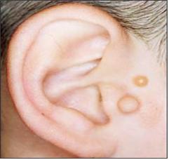
사진 26. 외이도 앞 부위에 난 두 개의 부이
Copyright ⓒ 2012 John Sangwon Lee, MD, FAAP
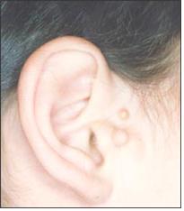
사진 27. 외이도 앞 부위에 난 두 개의 부이
Copyright ⓒ 2012 John Sangwon Lee, MD., FAAP
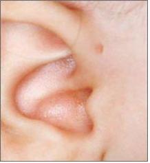
사진 28. 외이도 앞 부위에 난 이루공.
Copyright ⓒ 2012 John Sangwon Lee, MD., FAAP
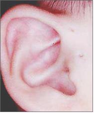
사진 29. 외이도 앞 부위에 난 두 개의 이루공
Copyright ⓒ 2012 John Sangwon Lee, MD., FAAP
|
다음은“이루공, 귀”에 관한 인터넷 소아청소년 건강상담 질의응답의 예 입니다. |
Q&A. 이루공– 귀에
Q.
- 안녕하세요..수고하십니다. 다름이 아니구요. 우리애기가요..지금백일이지나 5일째 구요, 남자아기구요..몸무게7.7kg이구요..그런데..38주에 제왕절개로 태어났습니다..만삭 때 낳으려고 했는데. 윤달이 있어..앞당겨서 낳았습니다.. 의사선생님께서는 38주 정도면 괜찮다고 해서 출산을 했는데요 잔병치레가 너무 많아요..후회가 되어..늘 애기한테 죄책감을 느껴요…다른 게 아니구요..
- 귀에요..양쪽 다요…구멍이 있어요..얼굴 볼쪽으로요…그게 태어나면서 가지고 나오는 거라고 하던데요.. 평생 그런 건가요? 그리고 그 부분에요..물 같은 게 나와요…심하면 수술해야 한다고 하는데요…저,, 너무 걱정이 되어 잠이 안 오거든요..다른 애들은 안 그런거 같은데..울고 싶어요..귀..구멍에 대해 설명 좀 해주세요…
A.
- 명자님
- 안녕하세요. 질문해 주셔 감사합니다.
- 나이, 성별, 과거, 현재, 가족의 병력, 진찰소견, 임상검사 결과 등 많은 정보가 있으면 더 좋은 답변을 드릴 수 있습니다. 주신 정보를 토대로 답변을 드립니다.
- 외이도 입구의 바로 앞 안면 피부에 바늘구멍만 한 구멍이 선천적으로 뚫려 있는 귀 기형을 이루공(사진 28, 29 참조)이라고 합니다.
- 이루공을 말씀하시는 것 같습니다.
- 거기에 염증이 생긴 것 같습니다.
- 아기엄마께서도 자상하고 아기에 대한 사랑을 많이 하시는 것 같습니다.
- 어떤 아이들은 키가 더 크고 어떤 아이들은 키가 더 작고 한쪽 팔이 없이 태어나기도 하고 심장 기형도 가지고 태어나기도 합니다.
- 이루공도 하나의 선천성 기형의 일종이지만 아주 미미한 선천성 기형 중 하나입니다.
- 엄마의 잘못으로 그런 기형이 생기는 것은 절대 아닙니다.
- 쉽게 치료 되니 걱정하시지 말고 적절히 치료를 해 주시면 될 것입니다.
- 소아청소년과에서 진찰 진단을 받으시고 그 문제에 관해 상담하시기 바랍니다.
- 귀에 생긴 여러 가지 기형–이루공 등을 참조하시기 바랍니다.
- 질문이 더 있으면 또 방문하세요. 감사합니다. 이상원 드림
|
다음은“이루공 유전”에 관한 인터넷 소아청소년 건강상담 질의응답의 예 입니다. |
Q&A. 이루공 유전에 대해서
Q.
- 안녕하세요..
- 먼저 질문에 대한 답변 감사드리구요, 한 가지 추가로 여쭤 볼께요..
- 귀기형(외이도 앞쪽에 난 하나의 이루공)은 유전이라고 하셨는데 아기 아빠랑 저는 둘 다 없는데 어떻게 아기한테 생길 수 있는 거죠?
A.
- 정님
- 안녕하세요. 질문해 주셔서 감사합니다. 좋은 질문입니다.
- 자녀의 나이 , 성별, 과거 병력, 가족 병력, 진찰소견, 임상검사 등의 정보를 많이 알수록 답변을 드리는데 도움이 됩니다. 주신 정보를 토대로 해서 답변을 드리겠습니다.
- 유전 인자로 유전됩니다.
- 혈우병과 같은 유전성 질환은 성염색체로 유전 되고 그 외 많은 다른 유전성 질환들은 상염색체로 유전되기도 합니다.
- 유전에는 우성으로 유전되기도 하고 열성으로 유전되기도 합니다.
- 상염색체로 유전되는 경우는 남아들이나 여아들에게 다 같은 빈도로 유전될 가능성이 있습니다.
- 유전학에서 표현률(Penetrance rate)이라는 말이 있다. 그 말은 유전병이 나타날 수 있는 빈도를 말합니다.
- 많은 유전성 질환이 불완전한 표현률로 나타나는 경우가 있습니다.
- 저는 유전학을 전문하지 안했습니다.
- 아기의 기형은 열성 표현 상염색체로 생긴 것 같습니다.
- 유전학 전문의나 소아청소년과 전문의 또는 이비인후과 전문의에게 문의하시는 것이 어떨까요.
- 도서관이나 서점에서 유전학 참고서를 참고하시는 것도 좋을 것입니다.
- 소아청소년과 청소년에서 진단 받으시고 이런 문제에 관해서 상담하시기 바랍니다.
- 질문이 더 있으면 다시 연락해 주시기 바랍니다. 감사합니다. 이상원 드림
|
다음은“귀에요… 부이 ”에 관한 인터넷 소아청소년 건강상담 질의응답의 예 입니다. |
Q&A. 귀에요… 부이
Q.
- 늦었지만 새해 복 많이 받으세요. *^^*
- 아이가 태어난 지 40여일 정도 되는데요.
- 태어나면서부터 귀 쪽에 이상한 것이 있어 궁금했는데 이곳 사이트에 들어와서 궁금증을 해소 했네요.
- “부이” 인 것 같거든요. 실로 묶어 두면 해결할 수 있다고 하셨는데요.
- 너무 어리지 않을까 해서요. 지금 해 주는 것이 좋을지 아니면 조금 더 자란 다음에 해 주는 것이 좋을지 시기를 알 수가 없어서 문의 드려요.
- 첫아이 때에는 그런 것이 없어서 몰랐는데 처음엔 너무 당황하고 속상해서 어떻게 해야 할지를 몰랐어요. 자세히 알려 주시면 감사하겠습니다.
A.
- 정원께
- 안녕하세요. 질문해 주셔서 감사합니다. 좋은 질문입니다.
- 자녀의 나이 , 성별, 과거 병력, 가족 병력, 진찰소견, 임상검사 등의 정보를 많이 알수록 답변을 드리는데 도움이 됩니다. 주신 정보를 토대로 해서 답변을 드리겠습니다.
- 부이의 크기, 수, 폭, 피부층에 접한 부이 뿌리의 크기, 자녀의 나이 등에 따라 치료를 다르게 할 수 있습니다.
- 어떤 부이는 상당히 크기 때문에 간단한 수술로 동네 소아청소년과에서 치료할 수 없습니다. 제가 실로 매어 치료한 부이 기형의 경우, 출생 후 바로 병원 신생아실에서 실로 매어 치료한 예입니다.
- 일반적으로 유치원에 들어갈 때쯤 부모도 자녀도 수술해 부이를 떼어 내고 싶으면 이비인후과에서 수술 치료를 받을 수 있습니다.
- 그 전에도 수술 치료를 받을 수 있지만 병원에 입원해야 할 경우도 있고 수술로 인한 세균 감염과 그로 인한 감염병이 생길 가능성이 더 있다는 것을 참작해서 좀 더 큰 후 수술 치료받는 것이 일반적입니다.
- 이 정도로 조그마한 선천성 기형은 생명에도 위험성이 없고 어떤 때는 이 기형으로 더 예쁘게 보일 수도 있고 그 아이 자신이 그것에 대해서 무관심하지만 부모들이 그것에 신경을 더 많이 쓰는 경우가 있습니다.
- 완전히 치료 받을 때까지 머리카락으로 부이 있는 그쪽을 가릴 수 있게 머리 손질을 적절히 해 주면 그런 귀 기형이 남들에게 잘 보이지 않을 수 있습니다.
- 소아청소년과에서 진찰 진단을 받으시고 이 문제에 대해서 상담하시기 바랍니다.
- 질문이 더 있으시면 다시 연락 주시기 바랍니다. 감사합니다. 이상원 드림
Congenital malformations of ears
Types of congenital ear malformations
1. Microtia- smaller ear. Congenital ear deformity with abnormally small pinna
2. Macrotia- Congenital anomaly of the ear with an abnormally large auricle
3. No auricle- Congenital ear malformation without auricle.
4. Auriclar skin tag- A skin pouch the size of a grain of rice, or a skin pouch the size of a pea in the area just in front of the entrance to the external ear canal. (See photo 2)
5. preauricular sinuses-An ear malformation with a congenital hole the size of a needle hole in the skin of the face in the area just before the entrance to the external auditory meatus (see photos 28 and 29).
6. Accessory ear
An ear malformation in which there is a congenital appearance of a prominent dermatophyte on the skin of the face in the region immediately anterior to the entrance to the external auditory meatus (see photos 22-27).
Buoys are the most common congenital ear anomaly.
7. Congenital anotia -An ear deformity that is congenitally absent from the outer and middle ear.
8. Protrude auricle, See photo 18.
9. Incomplete auricle, See photo 19.
10. loose auricle, See photo 20.
11. Darwin ear point on the auricle. See photo 21.
• Microtia: Underdeveloped outer ear
• Anotia: Missing one or both ears
• Protruding ears: More than 2 cm from the head
• Constricted ears (cup ears): Flattened or rolled outer ears
• Cryptotia: Upper ear underneath the scalp skin
• Stahl’s ear: Pointed outer ear
• Ear tag (accessory tragus): Cartilage bump in front of the ear
• Congenital earlobe deformities: Duplicate earlobes or earlobes with clefts or skin tags
• Ear hemangiomas: Small to large benign tumors
Treatment and Complications of Congenital Ear Anomalies
• If your child’s ear canal is infected with bacteria, it can become infected.
• Treat with appropriate antibiotics.
If the large ear can be surgically removed and treated.
• If the ear infection is not a major problem in terms of appearance, leave it as it is and conduct observational treatment.
• It is common to leaveCongenital auricular fistula as it is without any special treatment. Sometimes it can be infected.
• When the accessory ear is thin and elongated, it can be removed by tying the bottom of the accessory ear with a string (see photo 23).
• As can be seen in photo 23, the buoy is necrotic and the root of the accessory ear naturally falls off the skin after 1 to 2 weeks after holding the bottom of the accessory ear, which is thinly attached to the skin, with a thread. Otherwise, it can be removed by surgical excision.
• If the external or external auditory canal or middle ear is absent from birth, the external or external auditory meatus can be remodeled by plastic surgery.

Picture 18. Protruding auricle source; Used with permission from Mead Johnson Nutritional Division and Encyclopedia of Pediatric and Family Nursing

Photo 19. An imperfect auricle source; Used with permission from Mead Johnson Nutritional Division and Encyclopedia of Pediatric and Family Nursing

Photo 20. Loose auricle. Source; Used with permission from Mead Johnson Nutritional Division and Encyclopedia of Pediatric and Family Nursing

Picture 21. Darwin’s process on the auricle. Copyright ⓒ 2012 John Sangwon Lee, MD., FAAP

Photo 22. Accessory ear in front of the ear canal. Copyright ⓒ 2012 John Sangwon Lee, MD., FAAP

Photo 23. After 2 weeks of tying the bottom of the Accessory ear tightly with a thread, the Accessory ear may be necrotic and fall off. Copyright ⓒ 2012 John Sangwon Lee, MD., FAAP

Photo 24. Accessory ear in front of the ear canal. Copyright ⓒ 2012 John Sangwon Lee, MD., FAAP

Photo 25. Accessory ear in front of the ear canal. Copyright ⓒ 2012 John Sangwon Lee, MD., FAAP

Photo 26. Two Accessory ears in the anterior part of the ear canal. Copyright ⓒ 2012 John Sangwon Lee, MD, FAAP

Photo 27. Two Accessory ears in the anterior part of the ear canal. Copyright ⓒ 2012 John Sangwon Lee, MD., FAAP

Photo 28. Irugong in front of the external auditory meatus. Copyright ⓒ 2012 John Sangwon Lee, MD., FAAP

Picture 29. Two congenital auricular fistulae in the anterior part of the external auditory meatus Copyright ⓒ 2012 John Sangwon Lee, MD., FAAP
The following is an example of a Q&A for health counseling for children and adolescents on the Internet about “Irugong, Ear”.
Q&A. Irukong( Congenital auricular fistula) – in the ear
Q.
• Hello. Good job. It’s no different. It’s our baby.. It’s been 100 days or 5 days now, and it’s a baby boy..Weighs 7.7kg..But..was born by cesarean section at 38 weeks..I was going to give birth at full term. There is a leap month.. I gave birth earlier.. The doctor said it would be ok in 38 weeks, so I gave birth. There are too many minor illnesses.. I regret it.. I always feel guilty about my baby… It’s nothing else.
• Ears..Both…Have holes..Face toward the cheeks…They’re said to come with it at birth. Is it like that for the rest of your life? And in that part..Something like water comes out…If it’s severe, they say I need surgery…I’m so worried that I can’t sleep..I don’t think other kids are like that…I want to cry…Ears…holes Please explain about…
A.
• Myungja
• Good morning. Thank you for asking a question.
• We can give you a better answer if you have a lot of information such as age, gender, past, present, family history, examination findings, and clinical test results. We will respond based on the information you have provided.
• An ear deformity with a congenital hole the size of a needle hole in the skin of the face just in front of the entrance to the ear canal is called the preauricular sinus (see photos 28 and 29).
• You seem to be talking about Yiru Gong.
• There seems to be an inflammation there.
• It seems that the baby’s mother is also kind and loves her baby a lot.
• Some children are taller, some are shorter, some are born without an arm, and some are born with a heart defect.
• congenital auricular fistula is also a type of congenital anomaly, but it is one of the very minor congenital anomalies.
• It’s never the fault of her mother to have such a deformity.
• It can be easily treated, so don’t worry and treat it appropriately.
• Get a diagnosis from the Department of Pediatrics and consult about the problem.
• Please refer to the various deformities of the ear-perforation, etc.
• Come back with more questions.
Thank you. Lee Sang-won Dream The following is an example of a Q&A for health counseling for children and adolescents on the Internet about “Irugong genetics”. Q&A. About Irukong Oil Field
Q.
• Good morning.
• First of all, thank you for answering my question, and I have one more question. • You said that ear deformity (a single congenital auricular fistula in the front of the ear canal) is hereditary. A.
• Jung
• Good morning. Thanks for asking. That’s a good question.
• The more information you know about your child’s age, gender, past medical history, family history, examination findings, and clinical tests, the more helpful it is to give you an answer. We will give you an answer based on the information you provided.
• Inherited as a hereditary factor.
• Hereditary diseases, such as hemophilia, are inherited as sex chromosomes, and many other hereditary diseases are also inherited autosomal.
• Inheritance is either dominant or recessive.
• In the case of autosomal inheritance, it is likely that both boys and girls are inherited at the same frequency.
• There is a saying in genetics called the Penetrance rate. That means the frequency at which a hereditary disease can appear.
• Many hereditary disorders may present with an incomplete expression.
• I did not specialize in genetics.
• Your baby’s malformation appears to be caused by a recessive expressing autosomal.
• Why not ask your geneticist, pediatrician or otolaryngologist?
• You may want to consult a genetics reference book in your library or bookstore.
• Be diagnosed with children and adolescents and consult with them about these issues. • If you have more questions, please contact us again. Thank you. Lee Sang-won.MD
The following is an example of a Q&A on health counseling for children and adolescents on the Internet about “It is my ear…
Buoy “(Accessory ear)”.
Q&A. It’s an ear… buoy
Q.
• It’s late, but Happy New Year. *^^*
• It is about 40 days after the baby was born.
• Since I was born, I have been curious about something strange in my ears, but I came to this site and solved my doubts.
• It looks like a “buoy”. You said you could fix it by tying it up with a thread.
• I was afraid I might be too young. I don’t know when it’s better to do it now or if it’s better to do it after I grow up a little bit, so I’m asking.
• When I was my first child, I didn’t know because there was no such thing, but at first I was so embarrassed and upset that I didn’t know what to do. Any details would be appreciated. A.
•To the garden
• Good morning. Thanks for asking. That’s a good question.
• The more information you know about your child’s age, gender, past medical history, family history, examination findings, and clinical tests, the more helpful it is to give you an answer. We will give you an answer based on the information you provided.
• Treatment may vary depending on the size, number and width of the buoy, the size of the root of the buoy in contact with the skin layer, and the age of the child.
• Some buoys are quite large and cannot be treated with simple surgery at your local pediatric department. In the case of an accessory ear deformity that I treated with thread, it is an example that was treated by threading in the hospital neonatal room immediately after birth.
• In general, by the time kindergarten begins, both parents and children can have surgery to remove the buoy and receive surgical treatment at the otolaryngology department.
• You can receive surgical treatment before that, but you may need to be admitted to the hospital, and it is common to receive more surgery after surgery, taking into account the increased risk of bacterial infection and infection caused by surgery.
• A congenital anomaly this small is not life-threatening, and sometimes it can look prettier, and the child himself is indifferent to it, but parents pay more attention to it.
• Such ear deformities may be less visible to others if the hair is properly groomed to cover the buoy with the hair until fully healed.
• Get a diagnosis from the Department of Pediatrics and discuss this problem.
• If you have more questions, please feel free to contact us. Thank you. Lee Sang-won Dream
Abnormalities of the external ear[1]
Anotia/microtia
- Anotia is the total absence of the auricle, most often with narrowing or absence of the external auditory meatus.
- Strictly speaking, in microtia, there is some degree of malformation of the external ear (± narrowing or absence of the external auditory meatus) in contrast to a ‘small ear’ which is normally formed, as seen in Down’s syndrome.
- These conditions may be unilateral or bilateral – the latter is less common.
- Anotia is rare but seen in 20% of children with thalidomide-induced abnormalities. Microtia (along with protruding ears) is the most common ear problem encountered in plastic surgery. It is seen in 0.03% of all newborns. It is commonly associated with hemifacial microsomia.
- Between 6% and 16% of cases are associated with chromosomal abnormalities and up to 65% of cases occur in isolation.
- However, both can also be associated with first arch syndrome and the oculo-auricula-vertebral spectrum (of which Goldenhar’s syndrome is the most severe manifestation). Other associations include Treacher Collins’ syndrome, Nager and CHARGE syndrome (Coloboma, Heart defects, Atresia of the choanae, Restriction of growth and developmental delay, Genitourinary abnormalities, Ear anomalies), among others.
- External auditory meatus atresia must be ruled out early (within days in bilateral cases and months in unilateral cases), as delayed speech development can ensue.
- Surgical reconstruction of the auricle is usually carried out between 6-7 years of age.
- The options will involve either a reconstruction using rib cartilage (which requires several procedures, entails chest wall scarring and the ear does not grow with the child) or prosthetic reconstruction, which can achieve very lifelike results. The procedure can be done under local anesthetic but general anaesthetic is often preferred, particularly if the patient is a child.
- Postsurgical complications can include infection, hematoma and scarring. Psychological complications include disappointment due to unwarranted expectations – this can be obviated by careful counseling pre-operatively.
- The future promises exciting developments, including intrauterine diagnosis and treatment of severe ear deformities, gene therapy and tissue generation, in vitro cartilage growth, and the increasing use of prosthetic implants.
Macrotia
- A large but normally formed auricle is not usually associated with functional abnormality.
- It is defined as an ear that is two or more standard deviations from the mean. True macrotia is rare but may be seen in association with vascular malformations, hemihypertrophy, neurofibromatosis, and secondary to haemangioma.
- It is more conspicuous if the ear is prominent too.
- Surgical correction can be carried out. The Antia-Buch technique, which involves freeing the helical flap and repositioning it, is the most commonly used procedure.[4]
Preauricular accessory auricles
- These are usually found just anterior to the tragus and range from simple skin tags to complex structures containing cartilage.
- They are present in up to 1.5% of the population and, in isolation, usually present with no functional abnormality.
- They can be part of a syndrome (eg, Treacher Collins’ syndrome or Goldenhar’s syndrome).
- Simple lesions can be easily removed but, when they are more complex (eg, cartilage is involved), surgery is more tricky as all the cartilage needs to be removed and the superficially placed underlying facial nerve can be put at risk.
External auditory meatus atresia
- Congenital atresia of the external auditory canal is caused by a failure of canalization of the epithelial plug portion of the first branchial cleft.
- This results in the formation of a membranous or bony (or both) plate at the level of the tympanic membrane.
- There may be associated ossicular malformations. The stenosis may be for part or all of its length.
- Stenosis usually does not result in hearing loss if patency is maintained but atresia does.
- It is a rare condition (of the order of 1-5:200,000 live births) and is more common in boys. There is a positive family history in 14% of cases. Unilateral atresia is 3 to 6 times more likely to occur than bilateral atresia.
- External auditory meatus atresia is often associated with other abnormalities.
- Complications may include recurrent otitis media, cholesteatoma, and mastoiditis.
- Management is often multidisciplinary: ear, nose, and throat (ENT) surgeons will work alongside audiologists, plastic surgeons, pediatricians, and geneticists.
An audiological assessment is carried out in the first instance to rule out hearing impairment in the infant.
If it is unilateral, this should take place in the first few months of life but, if it is bilateral, within the first few days.[8]
Unilateral cases are usually managed well, although may have some trouble in localizing the exact direction of the sound. Problems arise if there is repeated otitis media or impacted cerumen in the normal ear.
In cases with bilateral abnormalities, bone conduction hearing aids are the first line of treatment (± followed by bone-anchored hearing aids).
This is eventually followed by surgery (after the age of 5 or 6 years), after CT assessment of the extent of the problem and regular audiological monitoring.
Surgery is always carried out after any correction of the auricle, as the unscarred skin is essential to the surgeon in fashioning a new auricle.
출처 및 참조문헌
- Nelson Textbook of Pediatrics 22ND Ed
- The Harriet Lane Handbook 22ND Ed
- Growth and development of the children
- www.drleepediatrics.com 제1권 소아청소년 응급 의료
- www.drleepediatrics.com 제2권 소아청소년 예방
- www.drleepediatrics.com 제3권 소아청소년 성장 발육 육아
- www.drleepediatrics.com 제4권 모유,모유수유, 이유
- www.drleepediatrics.com 제5권 인공영양, 우유, 이유식, 비타민, 미네랄, 단백질, 탄수화물, 지방
- www.drleepediatrics.com 제6권 신생아 성장 발육 육아 질병
- www.drleepediatrics.com제7권 소아청소년 감염병
- www.drleepediatrics.com제8권 소아청소년 호흡기 질환
- www.drleepediatrics.com제9권 소아청소년 소화기 질환
- www.drleepediatrics.com제10권. 소아청소년 신장 비뇨 생식기 질환
- www.drleepediatrics.com제11권. 소아청소년 심장 혈관계 질환
- www.drleepediatrics.com제12권. 소아청소년 신경 정신 질환, 행동 수면 문제
- www.drleepediatrics.com제13권. 소아청소년 혈액, 림프, 종양 질환
- www.drleepediatrics.com제14권. 소아청소년 내분비, 유전, 염색체, 대사, 희귀병
- www.drleepediatrics.com제15권. 소아청소년 알레르기, 자가 면역질환
- www.drleepediatrics.com제16권. 소아청소년 정형외과 질환
- www.drleepediatrics.com제17권. 소아청소년 피부 질환
- www.drleepediatrics.com제18권. 소아청소년 이비인후(귀 코 인두 후두) 질환
- www.drleepediatrics.com제19권. 소아청소년 안과 (눈)질환
- www.drleepediatrics.com 제20권 소아청소년 이 (치아)질환
- www.drleepediatrics.com 제21권 소아청소년 가정 학교 간호
- www.drleepediatrics.com 제22권 아들 딸 이렇게 사랑해 키우세요
- www.drleepediatrics.com 제23권 사춘기 아이들의 성장 발육 질병
- www.drleepediatrics.com 제24권 소아청소년 성교육
- www.drleepediatrics.com 제25권 임신, 분만, 출산, 신생아 돌보기
- Red book 29th-31st edition 2021
- Nelson Text Book of Pediatrics 19th- 21st Edition
- The Johns Hopkins Hospital, The Harriet Lane Handbook, 22nd edition
- 응급환자관리 정담미디어
-
소아가정간호백과–부모도 반의사가 되어야 한다, 이상원
-
Neonatal Resuscitation American heart Association
-
Neonatology Jeffrey J.Pomerance, C. Joan Richardson
-
Pediatric Resuscitation Pediatric Clinics of North America, Stephen M. Schexnayder, M.D.
-
Pediatric Critical Care, Pediatric Clinics of North America, James P. Orlowski, M.D.
-
Preparation for Birth. Beverly Savage and Dianna Smith
-
Infectious disease of children, Saul Krugman, Samuel L Katz, Ann A. Gershon, Catherine Wilfert
- 소아과학 대한교과서
- Other
|
Copyright ⓒ 2015 John Sangwon Lee, MD., FAAP 미국 소아과 전문의, 한국 소아청소년과 전문의 이상원 저“부모도 반의사가 되어야 한다”-내용은 여러분들의 의사로부터 얻은 정보와 진료를 대신할 수 없습니다. “The information contained in this publication should not be used as a substitute for the medical care and advice of your doctor. There may be variations in treatment that your doctor may recommend based on individual facts and circumstances. “Parental education is the best medicine.” |