선천성 담관 폐쇄로 생기는 신생아 황달(선천성 담관 폐쇄증으로 인한 황달), Neonatal jaundice due to bile duct obstruction
- 간에서 분비된 담즙이 십이지장관 속으로 흐르는 관을 담관이라고 한다.
- 담관은 간에서 시작해서 십이지장 관으로 연결된다.
- 드물게 어떤 신생아의 담관이 선천성으로 완전히 막힐 수 있다.
- 이렇게 선천성으로 완전히 막힌 담관을 선천성 담관 폐쇄라고 한다.
- 담관이 완전히 막혔을 때는 간에서 만들어진 담즙이 십이지장관 속으로 흘러 내려갈 수가 없다.
- 간에서 분비되는 담즙이 막힌 담관 부분까지 흘러 내려갔다가 막힌 담관 부분 이하로 더 이상 내려가지 않고 그 대신 그 담즙이 역류돼서 간 속으로 들어가 결국에는 핏속으로 흡수된다.
- 핏속으로 흡수된 담즙으로 인해 황달이 생긴다.
- 선천성 담관 폐쇄가 생기는 원인은 아직도 확실히 모른다.
- 출처와 참조문헌 –[부모도 반의사가 되어야 한다– 소아가정간호백과]-제 5권 인공영양, 비타민, 이유식–비타민 A 걸핍증, 신생아 황달 참조.
간내 담관 폐쇄증으로 인한 황달 참조.
- 선천성 담도 폐쇄로 생기는 신생아 황달(선천성 담도 폐쇄증으로 인한 황달)의 증상 징후
- 막힌 담관의 부위, 막힌 정도, 담관이 막힌 후 경과된 기간 등에 따라 증상 징후가 다르다.
- 전형적 선천성 담관 폐쇄증의 증상 징후는 대략 다음과 같다.
- 보통 생후 처음 몇 주 동안은 아무 증상 징후가 없을 수 있다.
- 생후 3~8주 정도 지나면 눈 흰자위와 피부에 황달기가 현저하게 나타나기 시작하고 피부와 눈이 노랗고 간이 붓고 커질 수 있다.
- 선천성 담관 폐쇄를 조기에 적절히 치료해 주지 않고 오랫동안 방치하면 황달은 점점 더 심해진다.
- 이 때 핏 속에 직접형 빌리루빈과 간접형 빌리루빈의 농도가 동시 증가된다.
- 이 병만 있을 때는 핏속 간접형 빌리루빈의 농도가 20mg/dl 이상으로 증가되지 않는 것이 통예이다.
- 시간이 더 경과돼서 병이 계속 진행되면 소화 장애, 혈액응고 장애, 성장 장애 등이 현저히 나타날 수 있다.
- 소변색이 노랗고 대변색이 백토 색과 같이 회백색이 될 수 있다.
- 담관이 완전히 폐쇄됐을 때는 수술로 새 담관을 만들어 주는 수술치료를 하거나 간 이식 수술을 해 주지 않으면 생후 1년 이내 사망하는 것이 보통이다.
■ 선천성 담관 폐쇄로 생기는 신생아 황달(선천성 담관 폐쇄증으로 인한 황달)의 진단
- 병력, 증상 징후, 진찰소견 등을 종합해서 이 병이 의심되면 핏속에 있는 직접형 빌리루빈과 간접형 빌리루빈의 농도, 간 기능검사, 간과 담관 초음파 사진검사, 간과 담관 CT스캔 검사 등으로 이 병을 확진할 수 있다.
- 담관 초음파 검사에 삼각대 징후(Triangular cord sign)가 있으면 이 병을 진단하는 데 많은 도움이 된다고 한다.
■ 선천성 담관 폐쇄로 생기는 신생아 황달(선천성 담관 폐쇄증으로 인한 황달) 치료
- 증상 징후, 담관 폐쇄의 정도, 합병증의 유무 등에 따라 치료할 수 있다.
- 담관 일부가 막혔는지, 전부 막혔는지, 완전히 또는 불완전하게 막혔는지에 따라, 그리고 막히지 않고 정상적으로 남아 있는 담관의 길이가 얼마나 되는지에 등에 따라, 간에서 나온 막히지 않은 담관을 십이지장에 연결시켜 주는 수술 치료를 한다.
이런 수술 치료를 할 수 없을 때는 다른 사람의 간을 이식해서 치료하기도 한다.
- 간에서 분비된 담즙이 십이지장관 속으로 흐르는 관을 담관이라고 한다.
- 담관은 간에서 시작해서 십이지장 관으로 연결된다.
- 드물게 어떤 신생아의 담관이 선천성으로 완전히 막힐 수 있다.
- 이렇게 선천성으로 완전히 막힌 담관을 선천성 담관 폐쇄라고 한다.
- 담관이 완전히 막혔을 때는 간에서 만들어진 담즙이 십이지장관 속으로 흘러 내려갈 수가 없다.
- 간에서 분비되는 담즙이 막힌 담관 부분까지 흘러 내려갔다가 막힌 담관 부분 이하로 더 이상 내려가지 않고 그 대신 그 담즙이 역류돼서 간 속으로 들어가 결국에는 핏속으로 흡수된다.
- 핏속으로 흡수된 담즙으로 인해 황달이 생긴다.
- 선천성 담관 폐쇄가 생기는 원인은 아직도 확실히 모른다.
출처와 참조문헌
- [부모도 반의사가 되어야 한다- 소아가정간호백과]-제 5권 인공영양, 우유, 이유, 비타민, 미네랄, 단백질, 탄수화물 지빙-비타민 A 걸핍증, 신생아 황달 참조.
- 간내 담관 폐쇄증으로 인한 황달 참조.
| 선천성 담도 폐쇄로 생기는 신생아 황달(선천성 담도 폐쇄증으로 인한 황달)의 증상 징후 |

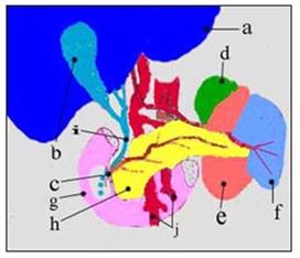
그림 135. 총수 담관(담도)이 선천성으로 막히면 신생아에게 황달이 생길 수 있다.
a-간, b-담낭, c-십이지장 유두(담즙과 췌장액이 분비되는 출구), d-부신, 선천성 담도 폐쇄증 e-좌 신장, f-지라, g-십이지장, h-췌장, i-총수 담관, j-혈관
Copyright ⓒ 2012 John Sangwon Lee, MD., FAAP
- 막힌 담관의 부위, 막힌 정도, 담관이 막힌 후 경과된 기간 등에 따라 증상 징후가 다르다.
- 전형적 선천성 담관 폐쇄증의 증상 징후는 대략 다음과 같다.
- 보통 생후 처음 몇 주 동안은 아무 증상 징후가 없을 수 있다.
- 생후 3~8주 정도 지나면 눈 흰자위와 피부에 황달기가 현저하게 나타나기 시작하고 피부와 눈이 노랗고 간이 붓고 커질 수 있다.
- 선천성 담관 폐쇄를 조기에 적절히 치료해 주지 않고 오랫동안 방치하면 황달은 점점 더 심해진다.
- 이 때 핏 속에 직접형 빌리루빈과 간접형 빌리루빈의 농도가 동시 증가된다.
- 이 병만 있을 때는 핏속 간접형 빌리루빈의 농도가 20mg/dl 이상으로 증가되지 않는 것이 통예이다.
- 시간이 더 경과돼서 병이 계속 진행되면 소화 장애, 혈액응고 장애, 성장 장애 등이 현저히 나타날 수 있다.
- 소변색이 노랗고 대변색이 백토 색과 같이 회백색이 될 수 있다.
- 담관이 완전히 폐쇄됐을 때는 수술로 새 담관을 만들어 주는 수술치료를 하거나 간 이식 수술을 해 주지 않으면 생후 1년 이내 사망하는 것이 보통이다.
| 선천성 담관 폐쇄로 생기는 신생아 황달(선천성 담관 폐쇄증으로인한 황달)의 진단 |
- 병력, 증상 징후, 진찰소견 등을 종합해서 이 병이 의심되면 핏속에 있는 직접형 빌리루빈과 간접형 빌리루빈의 농도, 간 기능검사, 간과 담관 초음파 사진검사, 간과 담관 CT스캔 검사 등으로 이 병을 확진할 수 있다.
- 담관 초음파 검사에 삼각대 징후(Triangular cord sign)가 있으면 이 병을 진단하는 데 많은 도움이 된다고 한다.
| 선천성 담관 폐쇄로 생기는 신생아 황달(선천성 담관 폐쇄증으로인한 황달) 치료 |
- 증상 징후, 담관 폐쇄의 정도, 합병증의 유무 등에 따라 치료할 수 있다.
- 담관 일부가 막혔는지, 전부 막혔는지, 완전히 또는 불완전하게 막혔는지에 따라, 그리고 막히지 않고 정상적으로 남아 있는 담관의 길이가 얼마나 되는지에 등에 따라, 간에서 나온 막히지 않은 담관을 십이지장에 연결시켜 주는 수술 치료를 한다.
- 이런 수술 치료를 할 수 없을 때는 다른 사람의 간을 이식해서 치료하기도 한다.
Neonatal jaundice due to bile duct obstruction
• The duct through which bile secreted from the liver flows into the duodenum is called the bile duct.
• The bile duct starts in the liver and connects to the duodenal lumen.
• Rarely, the bile ducts in some newborns can be completely blocked congenitally.
• This congenital blockage of the bile duct is called congenital bile duct obstruction.
• When the bile duct is completely blocked, the bile produced by the liver cannot flow down into the duodenum.
• Bile secreted from the liver flows down to the clogged bile duct and does not descend below the clogged bile duct. Instead, the bile refluxes into the liver and is eventually absorbed into the blood.
• Jaundice is caused by the absorption of bile into the blood.
• The cause of congenital bile duct obstruction is still unknown.
• Sources and References – www.drleepediatrics.com – Volume 5 Artificial Nutrition, Vitamins, Baby Food – Vitamin A Deficiency, Neonatal Jaundice See jaundice due to intrahepatic bile duct obstruction.
Signs, symptoms of neonatal jaundice due to congenital biliary atresia (jaundice due to congenital biliary atresia)
• Symptoms vary depending on the site of the blockage, the degree of blockage, and the elapsed time since the blockage of the bile duct.
• Symptoms of classic congenital biliary atresia include:
• Usually there may be no signs, symptoms during the first few weeks of life.
• After 3 to 8 weeks of age, jaundice begins to appear prominently on the whites of the eyes and skin, and the skin and eyes become yellow, and the liver may become swollen and enlarged.
• If congenital bile duct obstruction is left untreated for a long time without early and adequate treatment, jaundice will become progressively worse.
• At this time, the direct bilirubin and indirect bilirubin concentration in the blood increase simultaneously.
• In the presence of this disease, it is common practice that the concentration of indirect bilirubin in the blood does not increase to more than 20mg/dl.
• If the disease continues over time, digestive disorders, blood clotting disorders, and growth disorders may appear significantly.
• Your urine may be yellow and your stool may be grayish-white like clay.
• When the bile duct is completely blocked, it is common to die within 1 year of life unless surgery to create a new bile duct is performed or liver transplantation is not performed.
Diagnosis of neonatal jaundice (jaundice due to congenital biliary atresia) caused by congenital bile duct obstruction
• If the disease is suspected based on the medical history, symptom, signs, and examination findings, the disease can be diagnosed with the concentration of direct and indirect bilirubin in the blood, liver function test, liver and bile duct ultrasonography, and liver and bile duct CT scan. can be confirmed
• If there is a triangular cord sign-on bile duct ultrasonography, it is said to be very helpful in diagnosing this disease.
Treatment of neonatal jaundice caused by congenital biliary atresia (jaundice due to congenital biliary atresia)
• It can be treated depending on the symptoms, the degree of bile duct obstruction, and the presence or absence of complications.
• Surgical treatment to connect an unobstructed bile duct from the liver to the duodenum, depending on whether some, all, complete, or incomplete blockage of the bile duct, and how long the bile duct normally remains unblocked. When such surgical treatment is not possible, another person’s liver is transplanted for treatment.
• The duct through which bile secreted from the liver flows into the duodenum is called the bile duct.
• The bile duct starts in the liver and connects to the duodenal lumen.
• Rarely, the bile ducts in some newborns can be completely blocked congenitally.
• This congenital blockage of the bile duct is called congenital bile duct obstruction.
• When the bile duct is completely blocked, the bile produced by the liver cannot flow down into the duodenum.
• Bile secreted from the liver flows down to the clogged bile duct and does not descend below the clogged bile duct. Instead, the bile refluxes into the liver and is eventually absorbed into the blood.
• Jaundice is caused by the absorption of bile into the blood.
• The cause of congenital bile duct obstruction is still unknown.
Sources and References
• www.drleepediatrics.com-Volume 5 Artificial Nutrition, Milk, Weaning, Vitamins, Minerals, Protein, Carbohydrates-Vitamin A Deficiency, Neonatal Jaundice.
• See Jaundice due to intrahepatic bile duct obstruction.
Signs, symptoms of neonatal jaundice due to congenital biliary atresia (jaundice due to congenital biliary atresia)

Figure 135. Congenital blockage of the bile ducts (biliary ducts) can cause jaundice in newborns. a – liver, b – gallbladder, c – duodenal papilla (outlet of bile and pancreatic juice), d – adrenal gland, congenital biliary atresia e – left kidney, f – spleen, g – duodenum, h – pancreas, i – common bile duct, j – blood vessels. Copyright ⓒ 2012 John Sangwon Lee, MD., FAAP
• Symptoms vary depending on the site of the blockage, the degree of blockage, and the elapsed time since the blockage of the bile duct.
Symptoms of classic congenital biliary atresia include:
• Usually there may be no signs of symptoms during the first few weeks of life.
• After 3 to 8 weeks of age, jaundice begins to appear prominently on the whites of the eyes and skin, and the skin and eyes become yellow, and the liver may become swollen and enlarged.
• If congenital bile duct obstruction is left untreated for a long time without early and adequate treatment, jaundice will become progressively worse.
• At this time, the concentration of direct bilirubin and indirect bilirubin in the blood increases simultaneously.
• In the presence of this disease, it is common practice that the concentration of indirect bilirubin in the blood does not increase to more than 20mg/dl.
• If the disease continues over time, digestive disorders, blood clotting disorders, and growth disorders may appear significantly.
• Urine may be yellow and your stool may be grayish-white like clay.
• When the bile duct is completely blocked, it is common to die within 1 year of life unless surgery to create a new bile duct is performed or liver transplantation is not performed.
Diagnosis of neonatal jaundice due to congenital biliary atresia (jaundice due to congenital biliary atresia)
• If the disease is suspected based on the medical history, symptom signs, and examination findings, the disease can be diagnosed with the concentration of direct and indirect bilirubin in the blood, liver function test, liver and bile duct ultrasonography, and liver and bile duct CT scan. can be confirmed
• If there is a triangular cord sign-on bile duct ultrasonography, it is said to be very helpful in diagnosing this disease. Treatment of neonatal jaundice due to congenital biliary atresia (jaundice due to congenital biliary atresia)
• It can be treated depending on the symptoms, the degree of bile duct obstruction, and the presence or absence of complications.
• Surgical treatment to connect an unobstructed bile duct from the liver to the duodenum, depending on whether some, all, complete, or incomplete blockage of the bile duct, and how long the bile duct normally remains unblocked. do
• When such surgical treatment is not possible, another person’s liver is transplanted for treatment.
출처 및 참조문헌
- Nelson Textbook of Pediatrics 22ND Ed
- The Harriet Lane Handbook 22ND Ed
- Growth and development of the children
- www.drleepediatrics.com 제1권 소아청소년 응급 의료
- www.drleepediatrics.com 제2권 소아청소년 예방
- www.drleepediatrics.com 제3권 소아청소년 성장 발육 육아
- www.drleepediatrics.com 제4권 모유,모유수유, 이유
- www.drleepediatrics.com 제5권 인공영양, 우유, 이유식, 비타민, 미네랄, 단백질, 탄수화물, 지방
- www.drleepediatrics.com 제6권 신생아 성장 발육 육아 질병
- www.drleepediatrics.com제7권 소아청소년 감염병
- www.drleepediatrics.com제8권 소아청소년 호흡기 질환
- www.drleepediatrics.com제9권 소아청소년 소화기 질환
- www.drleepediatrics.com제10권. 소아청소년 신장 비뇨 생식기 질환
- www.drleepediatrics.com제11권. 소아청소년 심장 혈관계 질환
- www.drleepediatrics.com제12권. 소아청소년 신경 정신 질환, 행동 수면 문제
- www.drleepediatrics.com제13권. 소아청소년 혈액, 림프, 종양 질환
- www.drleepediatrics.com제14권. 소아청소년 내분비, 유전, 염색체, 대사, 희귀병
- www.drleepediatrics.com제15권. 소아청소년 알레르기, 자가 면역질환
- www.drleepediatrics.com제16권. 소아청소년 정형외과 질환
- www.drleepediatrics.com제17권. 소아청소년 피부 질환
- www.drleepediatrics.com제18권. 소아청소년 이비인후(귀 코 인두 후두) 질환
- www.drleepediatrics.com제19권. 소아청소년 안과 (눈)질환
- www.drleepediatrics.com 제20권 소아청소년 이 (치아)질환
- www.drleepediatrics.com 제21권 소아청소년 가정 학교 간호
- www.drleepediatrics.com 제22권 아들 딸 이렇게 사랑해 키우세요
- www.drleepediatrics.com 제23권 사춘기 아이들의 성장 발육 질병
- www.drleepediatrics.com 제24권 소아청소년 성교육
- www.drleepediatrics.com 제25권 임신, 분만, 출산, 신생아 돌보기
- Red book 29th-31st edition 2021
- Nelson Text Book of Pediatrics 19th- 21st Edition
- The Johns Hopkins Hospital, The Harriet Lane Handbook, 22nd edition
- 응급환자관리 정담미디어
-
소아가정간호백과–부모도 반의사가 되어야 한다, 이상원
-
Neonatal Resuscitation American heart Association
-
Neonatology Jeffrey J.Pomerance, C. Joan Richardson
-
Pediatric Resuscitation Pediatric Clinics of North America, Stephen M. Schexnayder, M.D.
-
Pediatric Critical Care, Pediatric Clinics of North America, James P. Orlowski, M.D.
-
Preparation for Birth. Beverly Savage and Dianna Smith
-
Infectious disease of children, Saul Krugman, Samuel L Katz, Ann A. Gershon, Catherine Wilfert
- 소아과학 대한교과서
- Growth and Development of Children, Eighth Edition, George H. Lowrey, Yearbook Medical Publishers
- Growth and Development of Children, Fifth Edition, E. H. Watson and G. H. Lowrey, Yearbook Medical Publishers
- Other
|
Copyright ⓒ 2015 John Sangwon Lee, MD., FAAP 미국 소아과 전문의, 한국 소아청소년과 전문의 이상원 저 “부모도 반의사가 되어야 한다”-내용은 여러분들의 의사로부터 얻은 정보와 진료를 대신할 수 없습니다. “The information contained in this publication should not be used as a substitute for the medical care and advice of your doctor. There may be variations in treatment that your doctor may recommend based on individual facts and circumstances. “Parental education is the best medicine.” |